16-year-Male presented with Swelling in right upper limb since 5 months, increasing in size since then, Episodes of high grade of fever and weakness in right upper limb
16-year-Male presented with Swelling in right upper limb since 5 months, increasing in size since then, Episodes of high grade of fever and weakness in right upper limb
No history of trauma
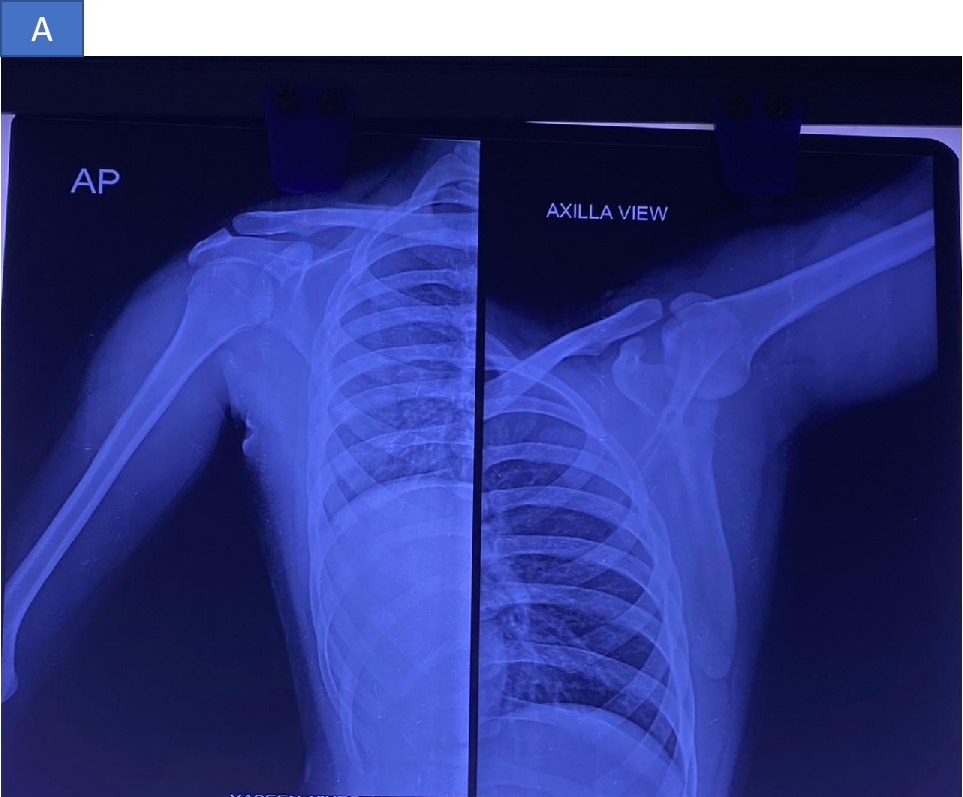
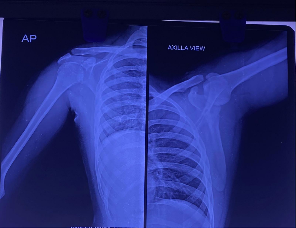
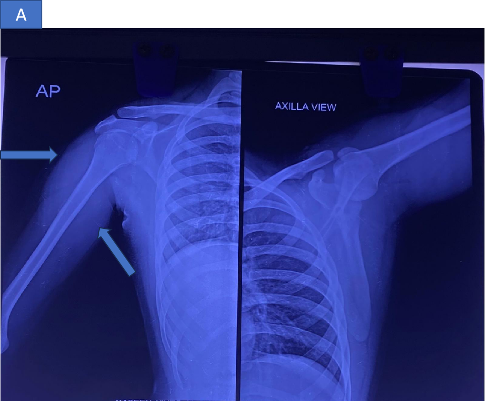
- X-ray showing prominent soft tissue shadow at the level of proximal 1/3rd humeral shaft region. No bony abnormality.
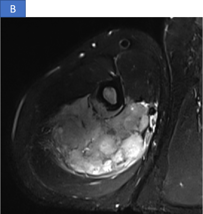
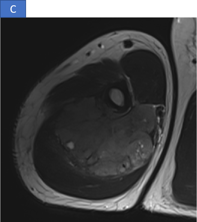
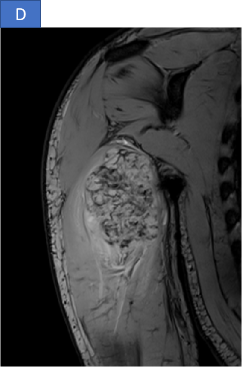
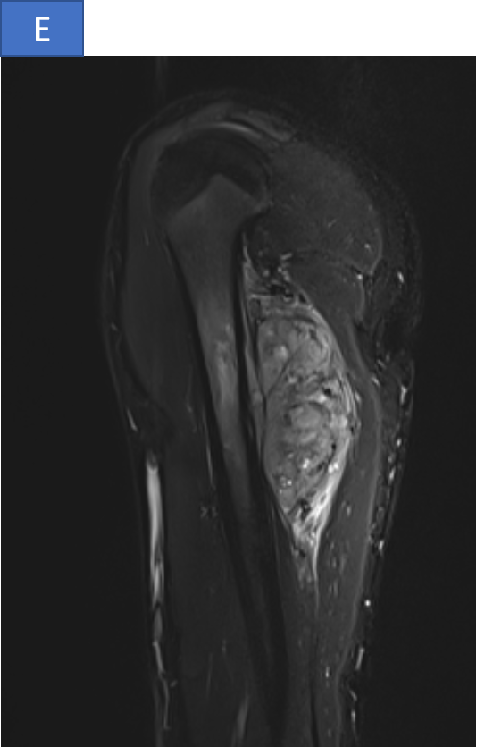
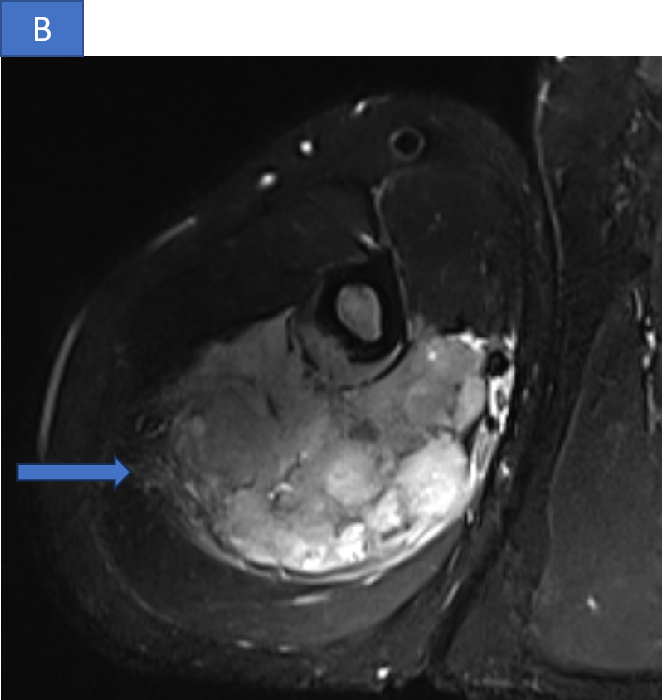
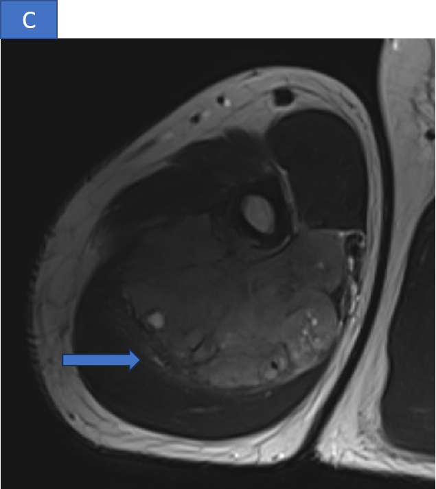
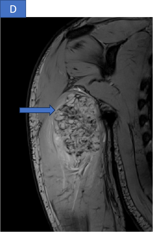
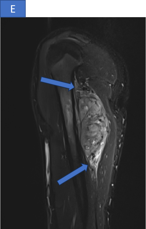
- A large altered signal intensity soft tissue lesion arising from the diaphysis of mid shaft region of humerus in posterior aspect with shallow surface cortical erosions, and spiculated periosteal reaction.
- The lesion demonstrates T2/STIR heterogeneous hyperintense signal and T1 hypointense signal with few cystic spaces. Multiple flow voids noted within lesion ( *). Mild intramedullary edema however no intramedullary extension.
DIAGNOSIS
- Periosteal osteosarcoma.
DISCUSSION
Parosteal osteosarcoma
- Rare, malignant, intermediate-grade, surface osteosarcomas that occur most commonly on the diaphysis of the femur and tibia.
- Second most common type of surface-based osteosarcoma after parosteal osteosarcoma.
- Account for 1-2% of all osteosarcomas.
- Periosteal osteosarcomas are seen in a wide age range from the first to the seventh decade with a peak in the second decade of life. Most common 15 to 25 years.
- The femur and tibia are the most common sites
- Patients typically present between the with regional pain and swelling.
Diagnostic criteria according to the WHO classification of soft tissue and bone tumors (5th edition):
- Imaging features of a bone tumor
- Histology of an intermediate-grade mostly chondroblastic osteosarcoma
- Origin from the surface of the bone under the periosteum
- Typically involves the diaphysis of long tubular bones
Radiographic features
- Broad-based cortically attached tumor with a partially mineralized soft tissue mass
- Extrinsic erosion of thickened underlying diaphyseal cortex with a surface-base crater
- Perpendicular or spiculated periosteal reaction
- Usually involves ~50% of the cortical circumference
- Intramedullary extension is rare
Radiographs shows a lesion that has a classic "sunburst or "hair on end" periosteal reaction.
- Treatment is usually neo-adjuvant chemotherapy, limb salvage surgical resection, followed by adjuvant chemotherapy.
- 20-35% chance of pulmonary metastasis
- Intermediate prognosis between parosteal and intramedullary osteosarcoma
Parosteal osteosarcoma
- Low grade
- Sclerotic lesion over the surface of bone.
- Thickening of the cortex and presence of a periosteal line between the tumor and the normal bone (string sign).
Periosteal osteosarcoma.
- Intermediate grade
- Broad based soft tissue mass
- Destruction of underlying bone with perpendicular periosteal reaction going into the soft tissue mass
References
- Bonar SFM, Klein MJ, O’Donell PG. Periosteal osteosarcoma. In: WHO Classification of Tumours Editorial Board. Soft tissue and bone tumours. Lyon (France): International Agency for Research on Cancer; 2020. (WHO classification of tumours series, 5th ed.; vol. 3). https://publications.iarc.fr
- MD/PhD, P. O. (n.d.). Periosteal osteosarcoma - Pathology - orthobullets. https://www.orthobullets.com/pathology/8016/periosteal-osteosarcoma
- Fox M & Trotta B. Osteosarcoma: Review of the Various Types with Emphasis on Recent Advancements in Imaging. Semin Musculoskelet Radiol. 2013;17(2):123-36. doi:10.1055/s-0033-1342969 - Pubmed
- Harper K, Sathiadoss P, Saifuddin A, Sheikh A. A Review of Imaging of Surface Sarcomas of Bone. Skeletal Radiol. 2021;50(1):9-28. doi:10.1007/s00256-020-03546-1 - Pubmed
- Murphey M, Jelinek J, Temple H, Flemming D, Gannon F. Imaging of Periosteal Osteosarcoma: Radiologic-Pathologic Comparison. Radiology. 2004;233(1):129-38. doi:10.1148/radiol.2331030326 - Pubmed
DR DEEPTI H V
Senior Consultant MHRG
DR ANKIT KATARIA
Cross-sectional fellow MHRG