
A 1-month-old male infant presented with complaints of midline swelling in the postero-superior region of head.
FINDINGS ON MRI BRAIN WITHOUT IV CONTRAST
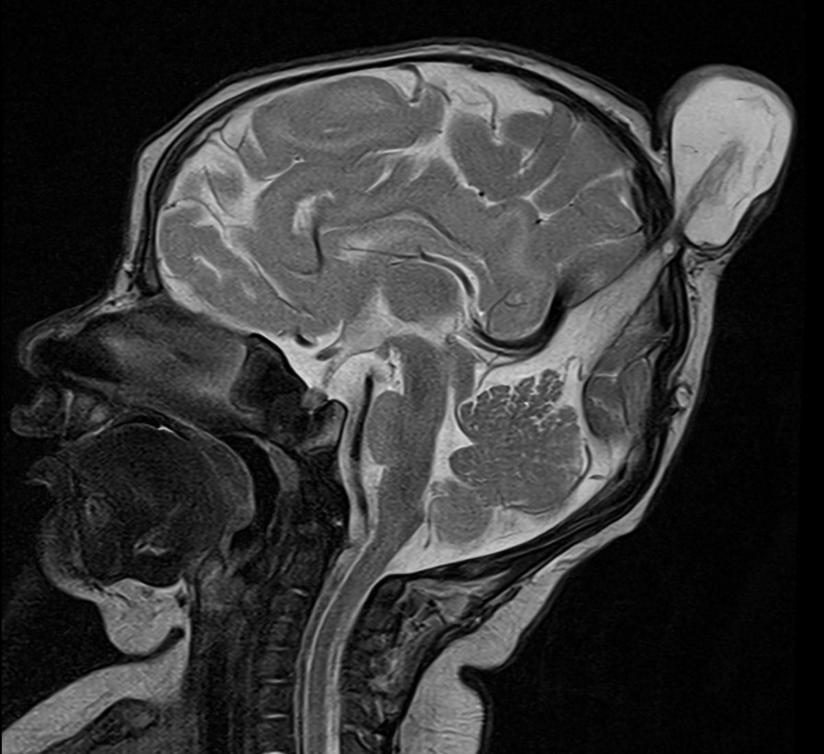
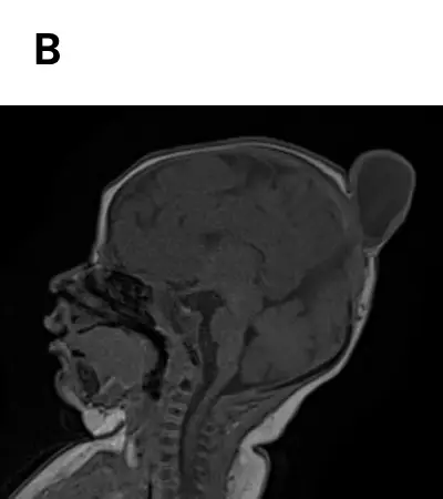
- A small well-defined cystic lesion seen in the midline parietal region appearing T2 hyperintense T1 hypointense (CSF signal intensity) with an intracranial extension and associated bone defect at the cranial vertex.
- CSF tract and primitive vertical falcine vein points towards the subcutaneous mass.
- Prominence of the superior sagittal sinus with superior peaking of posterior tentorium is seen giving spinning top configuration.
- Focal fenestration of the superior sagittal sinus.
- Fibrous stalk connecting the cephalocele.
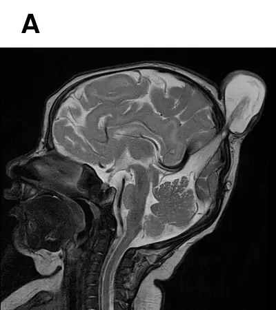
- Atretic parietal cephalocele
ATRETIC PARIETAL CEPHALOCELE :
- Atretic parietal cephaloceles, also known as atretic cephaloceles, are small subscalp lesions that consist of dura, fibrous tissue, and dysplastic brain tissue.
- Infants and young children.
- Palpable midline parietal soft tissue mass.
- Thought to represent involuted true cephalocele (meningocele or encephalocele) connected to the dura mater via a fibrous stalk.
- Increased incidence of intracranial anomalies.
IMAGING FEATURES:
- Subgaleal soft tissue mass with an intracranial extension via a sharply demarcated calvarial defect (cranium bifidum)
- CSF tract and vertical falcine vein point to the subcutaneous scalp mass
- Vertically oriented primitive falcine vein
- Fibrous stalk connecting the cephalocele
- Focal fenestration of superior sagittal sinus at the atretic parietal cephalocele
- Prominence of the superior cerebellar cistern and suprapineal recess
- Superior peaking of the posterior tentorium
- Spinning top configuration of the tentorial incisura
DIFFERENTIAL DIAGNOSES :
- Sinus pericranii
- Dermoid or epidermoid cyst
- Cephalohematoma
- Sebaceous cyst
- Vascular lesions (hemangioma)
References:
- Osborn AG, Salzman KL, Barkovich AJ. Diagnostic Imaging: Brain. Lippincott Williams & Wilkins. (2009) ISBN:1931884722.
- Wong SL, Law HL, Tan S. Atretic cephalocele - an uncommon cause of cystic scalp mass. Malays J Med Sci. 2012;17 (3): 61-3.
- Muralidharan CG, Aggarwal R, Singh D. Atretic parietal encephalocoele - An unusual diagnosis. Med J Armed Forces India. 2013;69 (1): 83-5.
Dr SRIRAM PATWARI
MBBS, MD, EDiNR
Senior Consultant Manipal Hospitals Radiology Group Yeshwantpur
Dr S Shreya
MBBS, MD
Cross section imaging fellow - MHRG